- Received January 26, 2023
- Accepted March 15, 2023
- Publication March 29, 2023
- Visibility 5 Views
- Downloads 0 Downloads
- DOI 10.18231/j.jdpo.2023.013
-
CrossMark
- Citation
Nevus lipomatosis cutaneous superficialis – A rare hamartoma
- Author Details:
-
Ekta Mishra
-
Deepika Sahu
-
Deepika Mishra
-
Shushruta Mohanty *
Introduction
Nevus lipomatosis cutaneous superficialis (NLCS) is an uncommon benign hamartomatous condition characterized by the presence of ectopic mature adiopocytes in the dermis. It was first reported by Hoffman and Zurhelle in 1921.[1] It has two clinical variants.[2], [3] The classical(multiple) form are present since birth or develop within third decade of life. It presents as groups of non-tender, soft, pedunculated, cerebriform, yellowish or skin-colored papules, nodules, or plaques in zosteriform, linear, or segmental distribution. Most common sites of predilection are pelvic girdle, lower trunk, gluteal region, and thigh.[4] On other hand Solitary variant of NLCS clinically mimicks a skin tag/fibroepithelial polyp and presents as a dome-shaped or sessile papule. It affects adults mostly in third to sixth decade. Other unusual sites reported in literatures are the scalp, eyelid, nose, and clitoris. [5] We here in report a case of classical form of Nevus lipomatosis superficialis in a 35 year old male.
Case Report
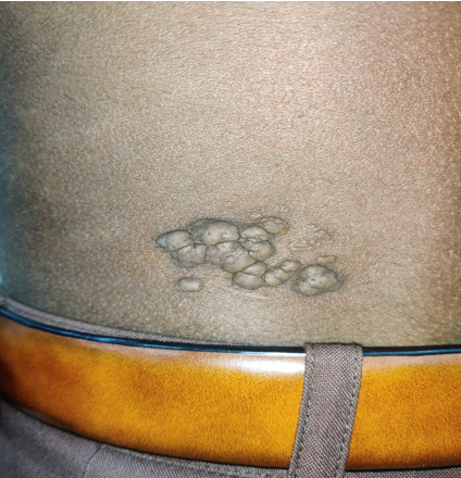
A 35 -year-old male presented with multiple nodules on left flank since 20 years. The lesion to start with presented as a solitary, painless swelling that was progressive in nature with no surface irregularity. Similar lesions in surrounding begin to appear which later coalased. Past medical or surgical history nor family history was significant. Clinical examination revealed multiple skin coloured soft nodular growths with comedo like plugs measuring 5 × 4 cm located over left lumbar area ([Figure 1]). The overlying skin was cerebriform and was non-ulcerated without any evidence of hypertrichosis, café-au lait macules or induration. Lymphnodes were not enlarged. No other significant lesions were detected in the body. Clinical differentials were plexiform neurofibroma and Nevus lipomatosus superficialis. All the routine laboratory investigations were within normal limits with viral markers (HBsAg/ HCV/ HIV) being non reactive. Punch biopsy was done under local anaesthesia and sample was sent for histopathological examination. Microscopy revealed epidermis lined by keratinised stratified squamous epithelium with hyperkeratosis and irregular acanthosis. The papillary dermis showed mature fat lobules admixed with collagen bundles. Reticular dermis was unremarkable. The adipocytes were closely associated with thin walled blood vessels and had no connection with underlying subcutis. ([Figure 2] a-b). This clinched our diagnosis as nevus lipomatosus cutaneous superficialis.


Discussion
NLCS usually presents as a developmental anomaly since birth or infancy where it is termed as nevus angiolipomatosis of Howell, but can also appear later in life. [5] It is usually asymptomatic, with neither sex predilection nor familial and is frequently devoid of any associated congenital defects.[5], [6] Other lesions known to coexist with NLS includes Cafe-au-lait macules, leukodermic spots, overlying hypertrichosis, and comedolike alterations.[5] Sometimes ulceration may occur following external trauma or ischemia. Another unusual presentation reported in the literature had rare features of recurrence associated with a foulsmelling discharge.[4] No such associated features were seen in our case apart from few comedo like plugs.
The precise etiopathogenesis of NLCS however remains unclear. There is no specific reason for predilection of lesions in the pelvic area. Deposition of adipose tissue in dermal connective tissue may be secondary to degenerative changes (metaplasia). The lesions could be explained by the possible origin of adipocytes from pericytes of dermal vessels, or developmental displacement of adipose tissue.[4], [6] NLCS may be a connective tissue nevus[7] and deletion of 2p24 in NLCS supports role of genetic factors in development of NCLS. [8]
Both the clinical variants i.e the classical and solitary types show similar histological features. The epidermis is normal to slightly attenuated.[4] Epidermal changes include mild to moderate acanthosis, basket weave hyperkeratosis, increased basal pigmentation, and focal elongation of rete pegs.[9] There is proliferation of mature adipocytes in the reticular dermis that may extend to papillary dermis with no connection with subcutaneous fat; for some authors, this is necessary for diagnosing NLCS.[6] The proportion of adipose tissue embedded between dermal collagen bundles is variable (10–50%).[5]
The clinical differential diagnoses are varied, that includes plexiform neurofibroma, smooth muscle hamartoma, nevus sebaceous, skin tag, and leiomyoma cutis.[10] These were ruled out histopathologically. Histological differentials include focal dermal hypoplasia (Goltz syndrome), lipofibromas, and giant acrochordons with fat herniation. Goltz syndrome is associated with ectodermal deformities, and exhibits almost entire replacement of the atrophic dermis by fat cells with the absence of collagen and skin appendages. Lipofibromas contain adipocytes, but lack dermal skin appendages. Skin tags show absence of adipocytes in dermis.[4], [5]
Conclusion
The treatment of choice of NLCS is surgical excision. However recurance following surgery is less with a single case reported in literature till date [6]. Larger lesions are treated with wide excision and skin grafting.[9] No malignant changes have been reported in association with NLCS until date. If left untreated, they can eventually increase in size causing cosmetic disfigurement. Histopathology plays a vital role in diagnosis of this entity.
Conflicts of Interest
None.
Source of Funding
None.
References
- E Hoffmann, E Zurhelle. Ubereinen nevus lipomatodes cutaneous superficialis der linkenglutaalgegend. Arch Dermatol Syph 1921. [Google Scholar]
- FB Yap. Nevus lipomatosus superficialis. Singapore Med J 2009. [Google Scholar]
- AC Buch, NK Panicker, PP Karve. Solitary nevus lipomatosus cutaneous superficialis. J Postgrad Med 2005. [Google Scholar]
- A Dhamija, A Meherda, P D'Souza, RS Meena. Nevus lipomatosus cutaneous superficialis: an unusual presentation. Indian Dermatol Online J 2012. [Google Scholar] [Crossref]
- SB Patil, S Narchal, M Paricharak, SS More. Nevus lipomatosus cutaneous superficialis: a rare case report. IJMS 2014. [Google Scholar]
- S Goucha, A Khaled, F Zéglaoui, S Rammeh, R Zermani, B Fazaa. Nevus lipomatosus cutaneous superficialis: report of eight cases. Dermatol Ther 2011. [Google Scholar]
- E Turan, Y Yesilova, D Ucmak, G Türkcü, OI Celik, MS Gürel. Nevus lipomatosus cutaneus superficialis associated with nevus sebaceous of Jadassohn. Indian J Dermatol Venereol Leprol 2014. [Google Scholar] [Crossref]
- N Cardot-Leccia, A Italiano, M C Monteil, E Basc, C Perrin, F Pedeutour. Naevus lipomatosus superficialis: A case report with a 2p24 deletion. Br J Dermatol 2007. [Google Scholar] [Crossref]
- S Dudani, A Col Malik, NS Brig Mani. Nevus lipomatosis cutaneous superficialis-a clinicopathologic study of the solitary type. Med J Armed Forces India 2016. [Google Scholar]
- P Bhushan, S S Thatte, A Singh. Nevus lipomatosus cutaneous superficialis: a report of two cases. Indian J Dermatol 2016. [Google Scholar] [Crossref]
